Franklin's Work On DNA
|
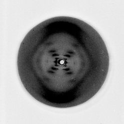
In 1950, Franklin was awarded a three-year Turner and Newall Fellowship to work in John T Randall's Biophysics Unit at King's College in London. She had been invited to work with a group of scientists studying living cells. She worked with a partner named Maurice Wilkins, and the two were assigned to work on DNA. There she made marked advances in x-ray diffraction techniques with DNA. These advances made it possible to extract finer DNA fibers, and Franklin was able to discover crucial keys to DNA's structure. Franklin discovered that DNA has two forms: a dry "A" form and a wet "B" form. Her conclusion of the wet form was probably helical in structure with phosphates on the outside of the ribose chains. Her mathematical analysis of the dry form, however, did not indicate a helical structure, so she spent over a year trying to resolve the differences. By early 1953, she had concluded that both forms had helical structures.
Working with Maurice Wilkins on the structure of DNA fibers, Franklin was in charge of the x-ray work. She decided that she could produce a controlled fashion of either of two forms (A or B) of DNA by varying the humidity of the fibers.Under agreements with Wilkins, however, Franklin could only work on the analysis of the A form. She gradually assembled enough data that would a double-helical structure, and was able to get two sets of high-resolution photos of crystallized DNA fibers. Franklin's work in x-ray crystallography produced the famous "Photo 51", which was used by Watson and Crick to help with their 3-Dimensional model of DNA. |
